Monday, November 14, 2011

Ex 8: Bone Classification, Structure & Relationships: An Overview

This lab is designed to provide students with an overview of bones through a variety of investigative activities.
https://sites.google.com/site/physiolablist/
The Objectives for this lab are:
  1. To list at least three functions of the skeletal system.
  2. To identify the four main kinds of bones.
  3. To identify surface bone markings and their function.
  4. To identify the major anatomical areas on a longitudinally cut long bone (or diagram of one).
  5. To identify the major regions and structures of an osteon in a histological specimen of compact bone (or diagram or model of one).
  6. To explain the role of the inorganic slats and organic matrix in providing flexibility and hardness to bone.


Hypotheses & Procedures
Part 1: Bone Markings & Classification.
     If students examine a variety of disarticulated bones then they will be able to classify them into one of the four anatomical groups: long, short, flat, or irregular and point out examples of bone markings.

Part 2: Gross Anatomy of the Typical Long Bone.
      If students examine a fresh cut bone as well as a cleaned dry bone that has been cut along its longitudinal axis, then they will be able to identify the major anatomical structures of a bone as an organ.


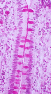
Part 3 Microscopic Structure of Compact Bone.
     If students observe a prepared slide of ground bone as well as a model of microscopic compact bone, then they will be able to identify the major regions and structures of an osteon.


Part 4: Chemical Composition of Bone:
     If students observe a sample of bone that has been baked then they will be able to identify the role of the organic compounds in bone because baking removes the collagen fibers of the matrix.
     If students observe a sample of bone that has been soaked in acid then they will be able to identify the role of the inorganic compounds in bone because acids dissolve the calcium salts.


Materials (on lab sheet)


Experiment/Data and lab questions

Part 1: Bone Markings & Classification.  
 
This is a picture of Cameron pointing to an irregular bone

This is a picture of Cameron pointing to the spongy, cancellous part of a long bone

 This is a picture of Cameron pointing to the sternum a flat bone.
 This is a picture of Cameron holding a long bone.
This is a picture of Cameron holding a short bone.
  Insert a picture of each type of bone, with a caption identifying its shape as long, short, flat, or irregular.  Within your caption, also identify any bone markings that show up in your picture.
Part 2: Gross Anatomy of the Typical Long Bone
      Insert at least one picture your partner pointing out a specific feature of long bone. Provide a caption to identify the specific feature.

Part 3 Microscopic Structure of Compact Bone
     Copy/paste this image into a draw program (like MS Paint).  Label the parts of the osteon that you can identify.  Save the image and use it to replace the unlabeled one into your blog, .


Part 4: Chemical Composition of Bone
Do treated bones retain the structure of untreated specimens?
no because they decay.

     Add a caption to this picture.  

Describe what happened to the matrix when the bone was baked.  How does this bone feel?
The bone became weak and brittle because they lack collagen

Describe what happened to the matrix when the bone was soaked in acid.  How does this bone feel?
the acid destroyed the periosteom making it feel softish


Optional: Include a video of your lab team demonstrating the effects of baking and acid on the matrix of bone.

Conclusion:
  1.  List at least three functions of the skeletal system.
  2. movement, protection, creating blood cells
  3. Identify the four main kinds of bones.
  4. long, short, irregular, flat
  5. Which surface bone markings and their function can you identify and describe?
  6. projections show where muscles are pulling on the bone.
  7. Which major anatomical areas on a longitudinally cut long bone (or diagram of one) can you identify?
  8. the cavity, the yellow bone marrow, spongy bone, compact bone.
  9. Which major regions and structures of an osteon in a histological specimen of compact bone (or diagram or model of one) can you identify?
  10. lacuna, matrix, central canal, lamella
  11. Explain the role of the inorganic slats and organic matrix in providing flexibility and hardness to bone.
  12. the slats and matrix help create the flexibility that is necessary for normal functions of the body

Monday, October 31, 2011

ex7: the skin & other body membranes

Ex7: The Skin & Other Body Membranes


I. Introduction/Background Information
        Refer to the Lab worksheet : https://sites.google.com/site/physiolablist/
There are several sections to this lab, generally designed to assist students in understanding the structures & functions of skin & other body membranes. The objectives of this lab are:
  1. to recount important functions of the skin/integumentary system
  2. to recognize and name the following structures from a slide, model or diagram
    • epidermis (including strata), 
    • dermis (papillary & reticular layers
    • hair follicles & hair
    • sebaceous glands & sweat glands
  3. to compare the properties of the dermis & epidermis
  4. to describe the distribution and function of the sebaceous glands, sweat glands, and hair 
  5. to differentiate between eccrine & apocrine sweat glands
  6. to ennumerate the factors determining skin color
  7. to describe the function of melanin
  8. to compare the structure and function of the major membrane types
  9. to list the general functions of each membrane type and note its location in the body
  10. to recognize by microscopic examination epidermal, mucous, and serous membranes
II. Hypothesis
Part 1: Basic structure of the skin: If students read about structures as they locate them on a skin model then they will be able to write out a response to each objective
Part 2: Appendages of the Skin: If students examine a prepared slide of human skin then they will be able to identify skin structures on a drawing.
Part 3: Plotting the Distribution of sweat glands: If bond paper is taped to skin coated with iodine and left for 20 minutes, then a distribution of sweat glands will be mapped out because the sweat glands will dissolve the iodine, which will then be absorbed by the paper.  As it reacts with the starch in the bond paper each sweat gland will produce a blue/black dot.
Part 4: Classification of Membranes: If students examine slides from trachea and small intestine, heart, as well as a freshly cut joint, then they can compare mucous, serous and synovial membranes.

III. Materials
    Model of Skin, prepared slides/pictures, iodine, 1cm x 1 cm bond paper squares, tape.

IV. Procedure
“See Lab Instructions” (you may include pictures of procedure)
Part 1: Basic structure of the skin: Compare the following structures identified in the pictures with the model in the classroom.
Skin slide
Human Skin Slide
Part 2: Appendages of the Skin:
Part 3: Plotting the Distribution of sweat glands: Insert a photo of each of your sweat distribution patches.  Label them "palm" and "forearm".
                                         palm                                      forearm
Part 4: Classification of Membranes:
pseudostratified ciliated columnar epithelium
mucosa of trachea


mucosa of small intestine


drawing of one villus
pericardium (around the heart)

V. Results/Observation/Data
List answers to questions from each section of the lab
Part 1: Basic structure of the skin: (No questions in this section)
Part 2: Appendages of the Skin:
wider view of the skin than the slides we used
Part i3: Plotting the Distribution of sweat glands:
the palm
Labeled  Pictures:
Part 4: Classification of Membranes:
small intestine is being used absorb while trachea is used to capture and move out of your body

yes because they have a bunch of nutrients around them


The pericardium is an example of a serous membrane.  It's too tiny to see, but the endothelium is composed of simple squamous epithelium.  List the specific names of the following serous membranes:
covering of the heart:
Lining of the cavity in which the heart resides:
Covering of the lungs:
Lining of the thoracic cavity:
Covering on the viscera:
Lining of the visceral cavity:
 

VI. Conclusion: Respond to each objective. What did you learn?
  1.  What are the important functions of the skin/integumentary system?
  2. Can you recognize and name the following structures from a slide, model or diagram?
    • epidermis (including strata), 
    • dermis (papillary & reticular layers
    • hair follicles & hair
    • sebaceous glands & sweat glands
  3. Compare the properties of the dermis & epidermis
  4. Describe the distribution and function of the sebaceous glands, sweat glands, and hair 
  5. Differentiate between eccrine & apocrine sweat glands
  6. Ennumerate the factors determining skin color
  7. Describe the function of melanin
  8. Compare the structure and function of the major membrane types
  9. List the general functions of each membrane type and note its location in the body
  10. Can you recognize by microscopic examination epidermal, mucous, and serous membranes?
  11.